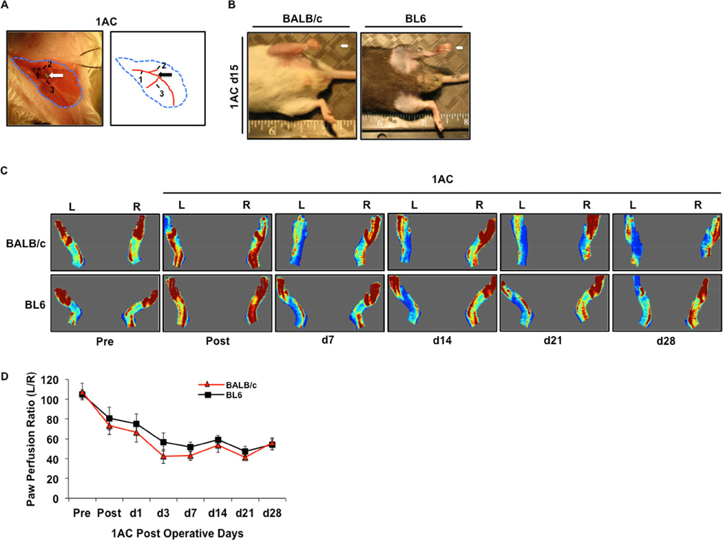
Figure 3. Comparison of BALB/c and BL6 pathology and blood flow after further refinement of subacute femoral artery occlusion by placement a single ameroid constrictor.
A, Representative image of a BALB/c mouse after placement of a single AC (1AC) on the proximal portion of the femoral artery, immediately proximal to the epigastric arterial branch. Arrow indicates site of constrictor placement. 1 Femoral artery. 2 Lateral Circumflex Femoral. 3 Superficial caudal epigastric artery. B, Representative gross anatomy photos and photomicrographs of TA muscle from BALB/c and BL6 mice subjected to subacute femoral artery occlusion via the placement of a single AC on the proximal femoral artery (1AC) for 15-days. C, Representative images from laser Doppler perfusion imaging in BALB/c and BL6 mice before and after 1AC placement up to 28 days post-operatively. D, Quantitation of paw perfusion in BALB/c (N=29) and BL6 (N=27) mice, represented as a ratio of ischemic (L) to non-ischemic (R) limb perfusion. * P<0.05 vs. day and ischemic model matched BL6.

