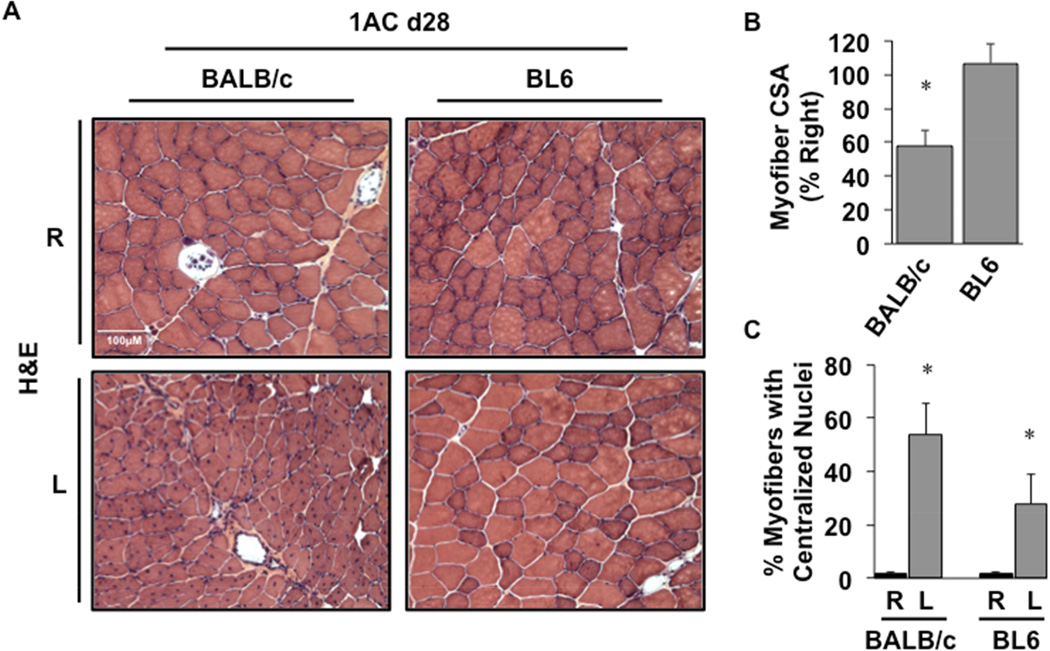
Figure 7. Ischemic skeletal muscle regeneration after sustained 1AC-mediated subacute ischemia.
A, Representative H&E-stained sections demonstrating TA morphology 28 days after 1AC placement. Sections of TA muscle were analyzed for mean myofiber CSA at day 28 and presented as a percentage of the right, non-surgical limb (B). *P<0.05 vs. BL6. C, The percentages of total myofibers with centralized nuclei. *P<0.05 vs. strain-specific contralateral (R) control.
All values are presented as means ± SEM.

