. 2015 Nov 27;4:e10616. doi: 10.7554/eLife.10616

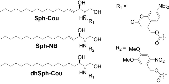
Figure 1. Structures of coumarin-caged sphingosine (Sph-Cou), nitrobenzyl-caged sphingosine (Sph-NB) and the negative control, coumarin-caged dihydrosphingosine (dhSph-Cou), respectively.

DOI: http://dx.doi.org/10.7554/eLife.10616.003

Figure 1.

Figure 1—figure supplement 1. Stability of caged Sph in cells.
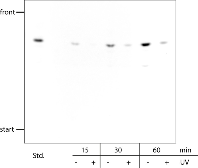
Figure 1—figure supplement 1.

HeLa cells were labeled with a pulse of 2 µM of Sph-Cou for the indicated times and then washed. Under +UV conditions, the cells were irradiated for 2 min. Cells were lysed, subjected to lipid extraction and separated by thin layer chromatography. The background was subtracted using Fiji software.
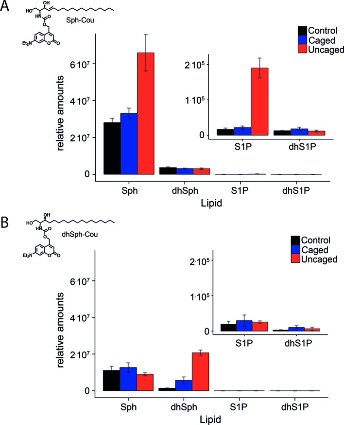
Figure 1—figure supplement 2. Comparative lipid analysis by mass spectrometry shows a successful uncaging reaction.

Figure 1—figure supplement 2.

Lipid extracts of HeLa cells pulsed with A) Sph-Cou and B) dhSph-Cou were AQC derivatized and measured on a TSQ Vantage mass spectrometer. Values are normalized to the C17 internal standards.