
Fig. 2.
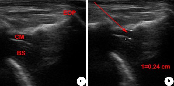
Measurement and observation of cisterna magna that is guided by ultrasound. a Cisterna magna and adjacent structures were displayed by ultrasound. b Depth of cisterna magna was measured at its optimal part. The puncture direction was designed based on the line from atlano-occipital fascia to the deepest part of cisterna magna. CM cisterna magna, BS brain stem, EOP external occipital protuberance