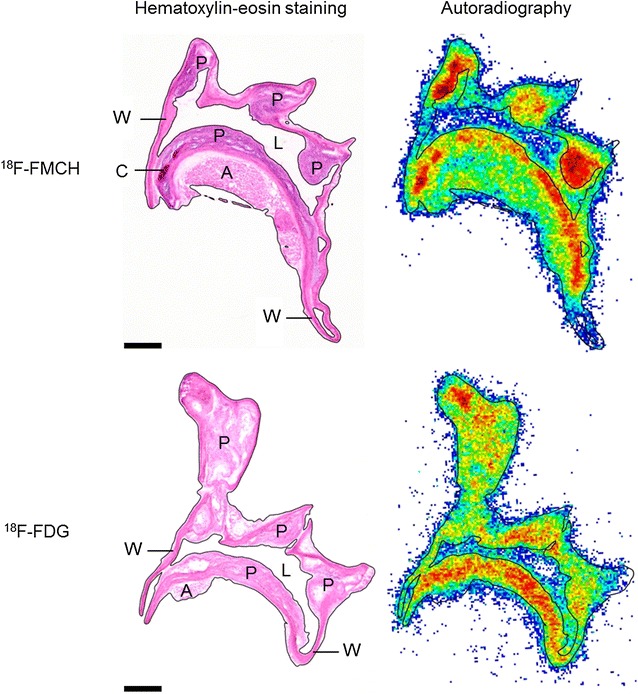
Fig. 3.

Histology and 18F-FMCH and 18F-FDG autoradiography of atherosclerotic aortas in diabetic IGF-II/LDLR−/−ApoB100/100 mice. Scale bar 500 µm. Both tracers show focal uptake in the atherosclerotic plaques (P) in the aortic arch and its branches. W normal vessel wall, A adventitia, C calcification, L lumen
