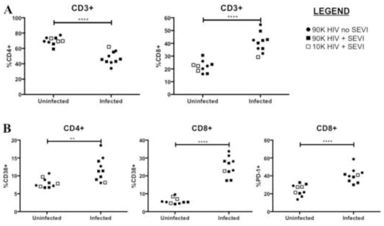
Figure 4. Immunophenotyping of peripheral blood lymphocytes at 4 weeks post-infection.
Mice were bled at 4wpi for analysis of HIV-associated pathogenic changes. Each point represents one mouse and the points are grouped according to HIV status. (A) Infected mice in all groups had significant decreases in CD4+ T cell proportions and increases in CD8+ T cell proportions compared to uninfected animals. (B) Infected mice had significant increases in proportions of activated CD4+ and CD8+ T cells as measured by CD38 expression. PD-1 is another measure of activation on CD8+ T cells and becomes chronically expressed in exhausted T cells. **P<0.01, ****P<0.0001, ns = not significant by unpaired t test.

