
FIGURE 3.
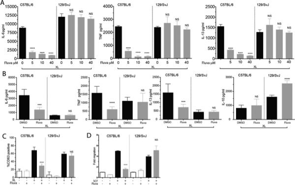
129/SvImJ mouse mast cells are resistant to fluvastatin. (A) C57BL/6J or 129/SvImJ BMMC were cultured in the indicated concentrations of fluvastatin and activated as described in Figure 1A. Culture supernatants were analyzed by ELISA. (B) Purified peritoneal mast cells from the indicated mouse strains were cultured for 24 hours with DMSO or 10μM fluvastatin for 24 hours, then activated with antigen. Culture supernatants were assessed by ELISA. (C) BMMC from the indicated strains were cultured as described in Figure 1A and activated with antigen for 1 hour prior to staining to detect surface CD63 by flow cytometry. (D) BMMC from the indicated strains were cultured with DMSO (-) or fluvastatin (10μM) for 24 hours, then tested for migration towards an SCF gradient as described in Materials and Methods. All data shown are from a minimum of three experiments. Data are means ± SEM of three independent experiments done in triplicate and analyzed by unpaired t-Test comparing fluvastatin- and DMSO (control)-treated groups. For each independent experiment n=3, (B) is representative of one of three independent experiments done in triplicate.