Fig. 1.
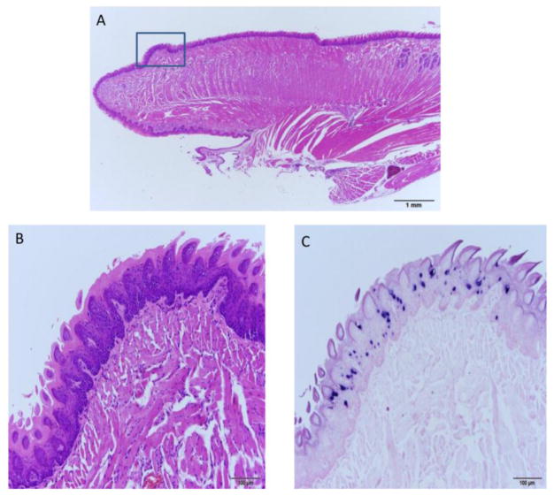
A–C The dorsal surface of the rostral tongue is susceptible to MmuPV1 infection. A) 10x sagittal section of the tongue of animal 3-3L sacrificed 3.5 months post infection and stained by H and E. The dorsal lesion on the rostral tongue is boxed. B) H and E (100x) and C) ISH (100x) of the boxed region in A showing a discrete focus of basal cell hyperplasia and strong in situ hybridization signal.

