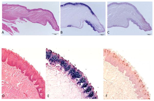
Fig. 2.
A–F By 8 months post infection both dorsal and ventral surfaces exhibit strong positivity for MmuPV1. A) Sagittal section of the tongue of mouse 3-3R sacrificed at 8 months post infection. H and E at 10x. B) ISH (20x) of the sagittal section showing strong ISH signal with clear demarcation of positive and negative sites. C) IHC(20x) of same section showing signal in the DNA-positive sites. D) H and E (100x), E) ISH (100x) and F) IHC (100x) illustrating the magnitude of viral infection.

