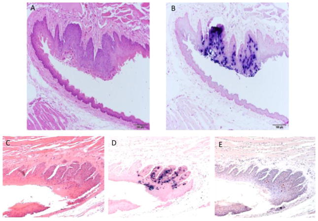
Fig. 6.
A–E Two lesions were found at the caudal ventral tongue. A) H and E (100x) of a sagittal/oblique section of the caudoventral tongue of animal 6-3R showing the area near the reflection to the sublingual oral mucosa. The animal was experimentally infected at several non-oral mucosal and cutaneous sites. B) ISH (100x) of an adjacent section showing strong staining near the reflection to the sublingual oral mucosa. C) H and E (100x) of a longitudinal parasagittal section of the tongue of animal 7-3L showing the reflection to the sublingual oral mucosa. The animal was originally infected at several non-oral mucosal and cutaneous sites. D) ISH (100x) of an adjacent section showing strong positivity at the reflection to the sublingual oral mucosa. E) IHC (100x) of an adjacent section showing capsid antigen presence in the same areas positive for MmuPV1 DNA.

