
Figure 2. Effect of SAMHD1 on lentiviral infection in dividing cells.
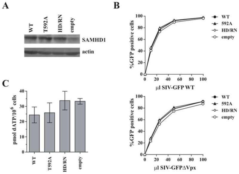
HeLa cells were transduced with lentiviral particles encoding either WT SAMHD1, the indicated SAMHD1 mutants, or an empty vector and selected with puromycin for 48 h. A) Total cell extracts were separated by SDS-PAGE and subjected to immunoblotting for SAMHD1 and actin as indicated. B) Cells were infected with increasing volumes of VSV-G-pseudotyped SIV-GFP (White et al., 2013a), with or without Vpx, and the percent infection (% GFP-positive cells) was determined by flow cytometry 48 h later. C) Cellular dNTPs were isolated at time of infection and the amount of dATP present per million cells was determined using a polymerase-based assay. Results in panels A and B are representative of at least 3 independent experiments. Error bars in panel C represent the mean and standard deviation of quantitation from at least 3 independently generated cell lines.