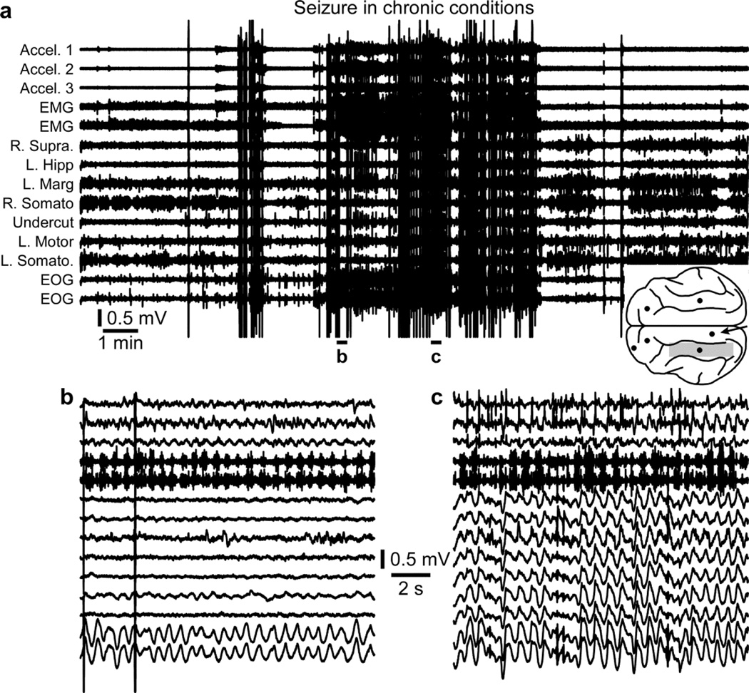
Figure 4. Seizure in chronic conditions.
a. Electrographic recordings of a seizure in chronic conditions. The location of recording electrodes and of the undercut region (shaded area) are indicated in the inset. b. Recordings from the accelerometer, EMG, and EOG shows rhythmic activities although none of the local field potential electrodes captured this rhythmic activity suggesting that the seizure was primarily focal. c. All recording electrodes display activities of the generalized seizure. b and c are enlarged segments from a as indicated.

