
Figure 1.
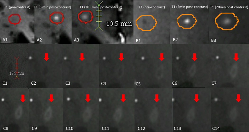
Dynamics of lesion enhancement. A, B: small, centrifugally enhancing lesions. A1, B1: T1-weighted images before contrast injection. A1: Isointense lesion. B1: Hypointense lesion. A2, B2: T1-weighted images 5min after contrast injection, demonstrating homogenously enhancing lesions. A3, B3: T1-weighted images 20min after contrast injection, demonstrating that the enhancement area has enlarged and that contrast material has spread out from the lesion center to the periphery. C: Centripetally enhancing lesion. C1: Precontrast dynamic-contrast enhanced T1-weighted images (DCE-MRI). C2-C14: Dynamic T1-weighted images, obtained using a different imaging sequence, at consecutive time points showing centripetal enhancement of the lesion. An initial ring of enhancement thickens over the time, eventually evolving into a homogenous, nodular enhancing lesion.