
Figure 2.
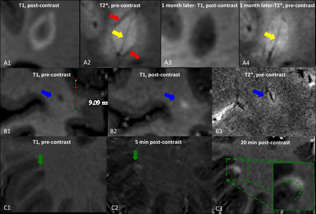
Susceptibility changes in active lesions and cortical enhancement. A: centripetally enhancing lesion, also shown in Figure 1C. A1, A3: T1-weighted images performed 5min after contrast injection. A1: the lesion shows ring enhancement. A3: One month later, enhancement has disappeared. A2, A4: T2*-weighted images. A2: The yellow arrow points to the hypointense central vein, and the red arrow shows a hypointense rim at the edge of the lesion. A4: One month later, the peripheral hypointense rim has almost disappeared, whereas the central vein persists. B1: T1-weighted images before contrast injection shows a small, hypointense lesion. B2: T1-weighted images 5min after contrast injection shows nodular enhancement of the lesion. B3: Precontrast T2*-weighted image shows a small vein centering the lesion. C: Leukocortical lesion. C1: Precontrast T1-weighted images shows a hypointense leukocortical lesion. C2: T1-weighted images 5min after contrast shows enhancement of the white matter portion of the lesion, but not the gray matter portion. C3: T1-weighted images 20min after contrast injection shows passage of contrast into the cortex.