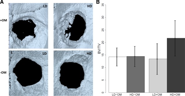
Figure 4.

Bone formation in critical‐size rat calvarial defects. A: Three‐dimensionally reconstructed high‐resolution μCT image of defects implanted with cells/scaffolds after 8 weeks of healing. Note the new bone formed in the four groups; with osteogenic medium (+OM) or without osteogenic medium (−OM) with different densities of cell seeding (low density (LD) or high density (HD)). B: Quantification of percentage of area and volume of bone regeneration in calvarial defects after 8 weeks of healing. Implantation of scaffolds containing cells seeded at high density and cultured with osteogenic medium exhibit a significant percentage of bone volume (p = 0.038). [Color figure can be viewed in the online issue, which is available at wileyonlinelibrary.com.]
