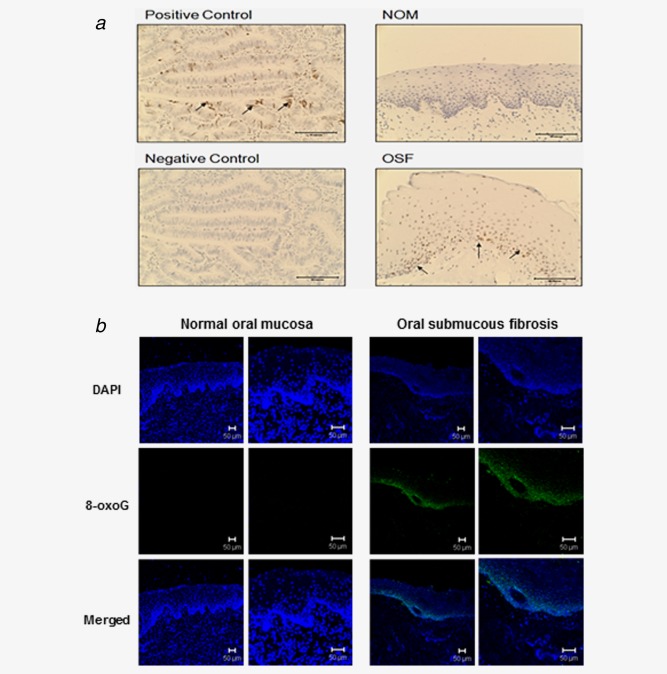
Figure 4.

DNA DSB and oxidative DNA damage were detected in OSF tissues (a) Immunohistochemical staining for phospho‐Histone H2A.X in OSF tissue. OSF tissue showed DNA damage foci in basal cell layer as compared to NOM. Arrows indicate positive cells. Positive control: human colon cancer. Representative photomicrographs are shown. Scale bar 50 µm. C: control (b) OSF tissues showed positive staining for 8‐oxoG FITC‐conjugate in basal cell area seen by confocal microscopy. Scale bar 50 µm. [Color figure can be viewed in the online issue, which is available at wileyonlinelibrary.com.]
