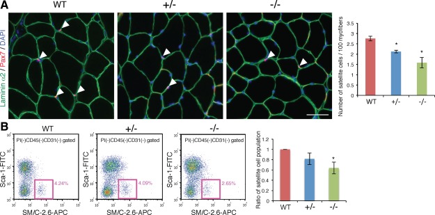
Figure 3.

Decrease of satellite cells in Ten‐4‐deficient mice. (A): Immunohistochemistry of Pax7 (red) in WT, Ten‐4+/−, and Ten‐4−/− TA muscle sections. Arrowheads indicate Pax7‐positive cells located between a myofiber and basement membrane labeled with laminin α2 staining (green). DAPI staining (blue) was used to visualize nuclei. Scale bar = 50 µm. The number of satellite cells per cross‐sectional 100 myofibers was counted. Error bars, SEM; *, p < .05. (B): Population of satellite cells from WT, Ten‐4+/−, and Ten‐4−/− TA muscles by flow cytometry. The CD31−CD45−Sca‐1−SM/C‐2.6+ cells were analyzed as muscle satellite cells using the MoFlo flow cytometer. Pink boxes represent the satellite cell population, and values in pink denote percentages of satellite cells in total mononuclear cells, except for CD45+ lymphocytes/leukocytes and CD31+ endothelial cells. The percentage of the WT satellite cell population was set as 1.0. Error bars, SEM; *, p < .05. Abbreviations: APC, allophycocyanin; TA, tibialis anterior; WT, wild type.
