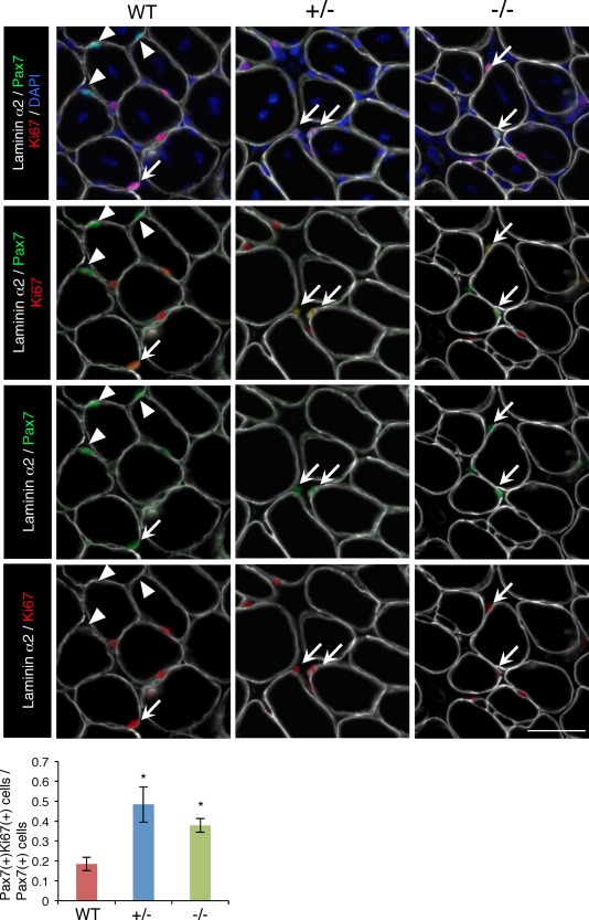
Figure 6.

Increased activated satellite cells in regenerated TA muscle from WT, Ten‐4+/−, and Ten‐4−/− mice. Immunostaining of laminin α2 (white), Pax7 (green), and Ki67 (red) in the TA muscle tissue of WT, Ten‐4+/−, and Ten‐4−/− mice, 7 days after CTX injection. Arrows indicate Pax7‐ and Ki67‐double positive cells, and arrowheads represent Pax7‐single positive cells. DAPI staining (blue) was used to visualize nuclei. The ratio of the numbers of Pax7‐ and Ki67‐dual positive cells/Pax7‐single positive cells was quantified. Error bars, SEM; *, p < .05. Scale bar = 50 µm. Abbreviation: CTX, cardiotoxin; TA, tibialis anterior; WT, wild type.
