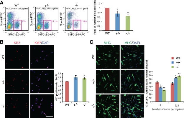
Figure 7.

Promoted cell proliferation and differentiation in the culture of purified satellite cells from Ten‐4‐deficient mice. (A): Flow cytometry profiles of satellite cells in WT, Ten‐4+/−, and Ten‐4−/− mouse skeletal muscles from fore‐limb and hind limbs. The CD31−CD45−Sca‐1−SM/C‐2.6+ cells were analyzed and sorted out as muscle satellite cells using the MoFlo flow cytometer. Pink boxes represent the satellite cell population, and values in pink denote percentages of satellite cells in total mononuclear cells, except for CD45+ lymphocytes/leukocytes and CD31+ endothelial cells. The percentage of WT satellite cells was set as 1.0. Error bars, SEM; *, p < .05; **, p < .01. (B): Immunostaining of Ki67 in WT, Ten‐4+/−, and Ten‐4−/− cells cultured for 3 days with the proliferation medium. Cells were stained with anti‐Ki67 antibody (red) and DAPI (blue) to label proliferative cells and nuclei, respectively. The relative numbers of proliferative cells were calculated. The cell number of the WT culture was set as 1.0. Error bars, SEM; *, p < .05; Scale bar = 100 µm. (C): Immunostaining of MHC in WT, Ten‐4+/−, and Ten‐4−/− cell cultures 2 days after induction of differentiation. Cells were stained with anti‐MHC antibody (green) and DAPI (blue) to visualize differentiating myoblasts/myotubes and nuclei, respectively. The number of nuclei in MHC‐positive myotubes or cells was measured and quantified. Error bars, SEM; *, p < .05; **, p < .01. Scale bar = 100 µm. Abbreviations: APC, allophycocyanin; MHC, myosin heavy chain; WT, wild type.
