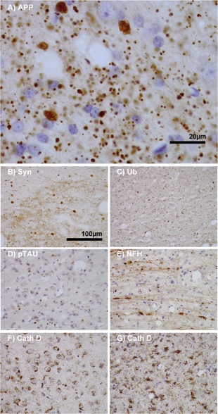
Figure 3.

Features of thalamic pathology. (A) High power (100× objective) photo‐micrographs of thalamic APP pathology shows large numbers of 1–2 μm diameter positive elements as well as smaller numbers of much larger spheroids (>6 μm). For comparative purposes, a number of other pathological features were examined for similarities and these were photographed at 40×. (B) Sy38 shows large spheroids of similar size to APP and significant overall loss, but no small deposits. (C) Ubiquitin shows large numbers of small ‘dot‐like’ deposits that are similar in size but very much fewer that those observed with APP. (D) Phospho tau (AT8) labelling shows frequent apparently neuritic labelling. (E) SMI‐32, neurofilament H labelling showed strong spheroid labelling in axons emerging from the medial lemniscus but much fewer spheroids in the grey matter. (F) Cathepsin D showed typical lysosomal labelling of small spherical structures in a perinuclear location, predominantly visible in neurons of the PO in NBH animals. (G) Cathepsin D in ME7 animals showed predominantly glial localization and consisted of larger structures or accumulations in perinuclear but also in some glial processes.
