Figure 8.

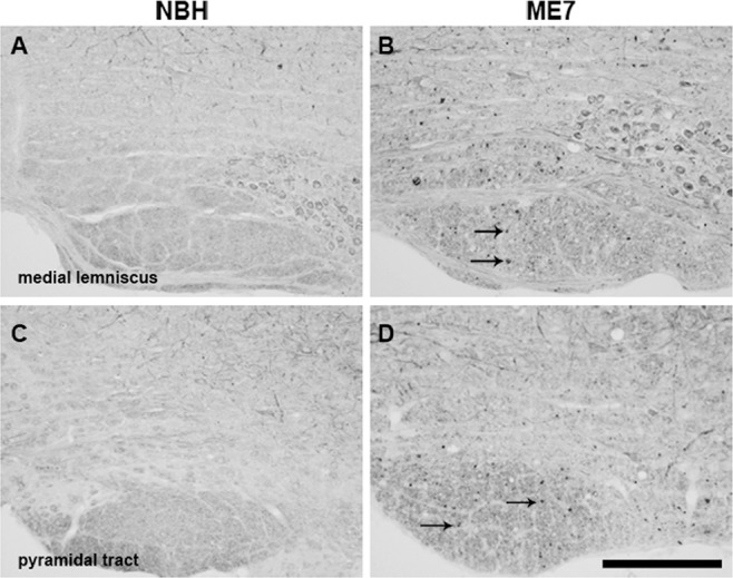
Axonal pathology in the medial lemniscus and pyramidal tract. Immunolabelling for SMI32 in coronal sections, at the rostrocaudal level, containing PrTN and Sp5I of experimental groups. High magnification images of medial lemniscus (B) and the pyramidal tract (D) show significant white matter pathology in ME7 animals, as indicated by intense SMI32 labelling (denoted by black arrows) that is not present in NBH (A,C). PrTN, principal trigeminal nucleus; Sp5I, spinal trigeminal nucleus, part interpolari. Scale bar = 200 μm.
