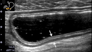
Figure 2.

Ultrasonography (US) images of the normal gastrointestinal (GI) tract wall. Five layers are visible in the normal stomach (arrow). The first layer corresponds to the border echo and a part of mucosa, the second layer is the rest of mucosa, the third layer is muscularis mucosa, submucosa, and a part of muscularis propria, the fourth layer is the rest of muscularis propria, and the fifth layer is serosa and border echo.
