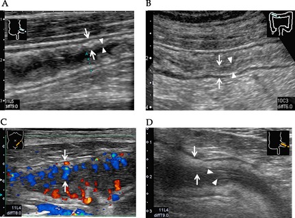
Figure 5.

Ultrasonography (US) grading of gastrointestinal graft‐versus‐host disease (GI‐GVHD). (A) US grade 1: Mucosa and submucosal layers are slightly thickened in the wall layer (arrow) and the boundary between internal low echoic layer (mucosa) and the third layer is clear (arrow head). (B) US grade 2: Diffuse wall thickness (arrow) and the boundary of internal low echoic layer and submucosa is obscure (arrow head). (C) US grade 3: Internal low echoic layer is markedly low, and increased Doppler signaling is seen in the layer (arrow). (D) US grade 4: Desquamated mucosal epithelium (arrow head) is seen in the internal lumen with wall thickening (arrow).
