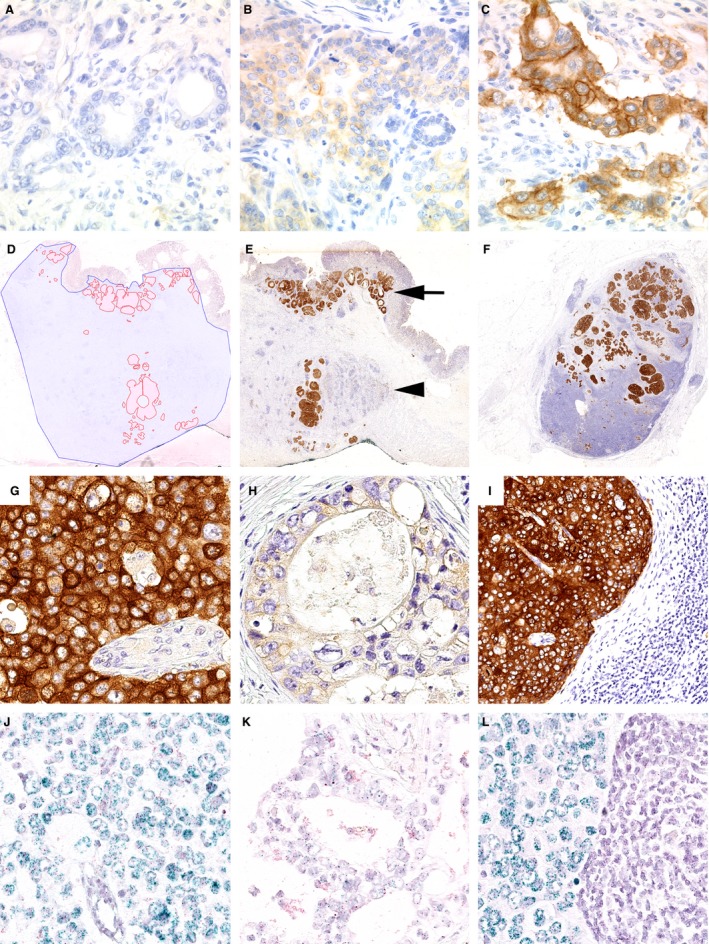
Figure 1.

Hepatocyte growth factor receptor (MET) protein expression in gastric cancer. The expression of MET was studied by immunohistochemistry (IHC) (A–C, E–I) and chromogenic in‐situ hybridization (D, J–L). The entire gastric cancer cohort was screened and three representative cases were selected for MET IHC 0 (A), 1+ (B), 2+ (C) and 3+ (G). Using a viewer program with a polygon line drawing function we traced manually the outlines of the total tumour tissue area (blue) and the MET‐amplified tumour area (red; D). This illustrates the heterogeneity of MET expression and MET amplification. Less than 10% of the entire tumour area showed MET‐IHC 3+ (E, G), while other areas were MET‐IHC 0 or 1+ (H). MET was amplified (J) or not (K). A corresponding lymph node metastasis of the same patient showed MET‐IHC 3+ (F, I) and MET amplification of the metastatic tumour cells (L). The spatial distribution of MET status‐positive tumour cell clones is also illustrated in (E). Immunostaining was localized at the cell membrane as well as in the cytoplasm. MET‐positive tumour cell clones were found near the mucosal surface (arrow) and in the tumour centre (arrowhead; E).
