
Figure 4.
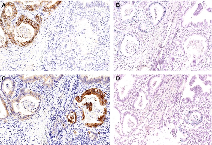
HER2/neu‐ and hepatocyte growth factor receptor (MET) overexpression in a single patient. A single patient showed overexpression of HER2/neu (A, B) and MET (C, D) in spatially distinct tumour areas. Four serial sections were stained immunohistochemically (A, C) and by chromogenic in‐situ hybridization (B, D). Note the distinct immunoreactions of HER2/neu and MET. HER2/neu showed a characteristic delicate basolateral staining of the cell membrane, while MET immunostaining was also strong within the cytoplasm.