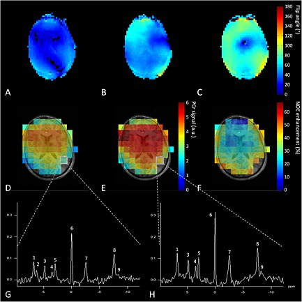
Figure 6.

(A–C) Results of in vivo experiment using the complete setup showing different B 1 + maps of the 1H field: (A) in CP− mode, (B) homogenized for the complete brain, and (C) locally optimized for occipital lobe. Showing the ability to use B 1 shimming with the coil setup. Background images were acquired with the 8‐CH 1H head coil driven with global B 1 optimization, showing fairly homogeneous field distribution. (D–F) 31P spins were excited and signal was acquired with the BC (D) without and (E) with NOE enhancement of PCr; (F) the global enhancement map. (G, H) An example of 31P spectra taken from the same voxel (G) without NOE enhancement and (H) with NOE enhancement. Spectra were obtained in 7 min 48 s with an approximate voxel size of 38 cm3. The metabolites present in these spectra are (1) phosphoethanolamine (PE), (2) phosphocholine (PC), (3) Pi, (4) glycerophosphoethanolamine (GPE), (5) glycerophosphocholine (GPC), (6) PCr, (7) γ‐ATP, (8) α‐ATP and (9) NADH.
