
Figure 2.
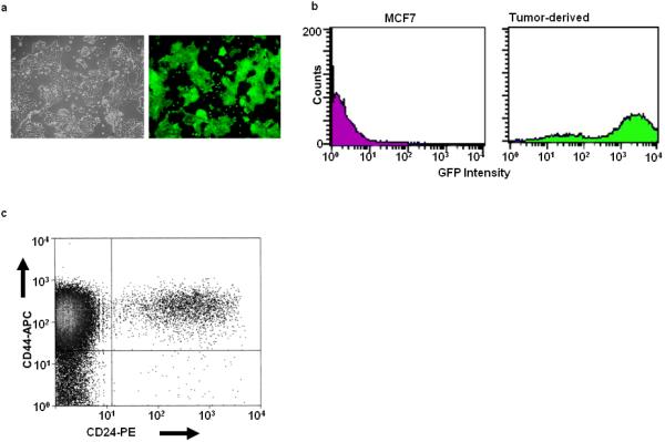
Phenotypic characterization of tumor-derived Oct3/4-GFP cells. Tumors arising in nu/nu mice inoculated with the CSC-like MCF7 cells shown in Fig. 1 were minced, dissociated with trypsin and collagenase, and propagated for 2-3 weeks in vitro in the presence of G418. (A): Visible light + UV low power microscopic views of a typical field showing GFP expression by the entire population of G418-resistant cells. (B): Flow cytometric evaluation of the re-cultured tumor-derived CSC-like cells confirming the expression of GFP by virtually the entire population (green curve). CD44hi/CD24lo-depleted cells were included as a negative control (magenta curve). (C): CD44/CD24 staining showing that the tumor-derived cells shown in (A) retained the CD44hi/CD24lo phenotype.