
Figure 4.
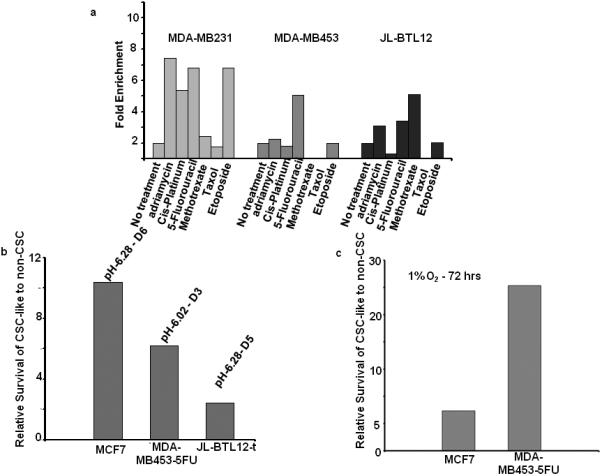
Differential sensitivities of CSC-like cells to chemotherapeutic drugs, acidosis, and hypoxia. Each of the above-described GFP+ CSC-like cell lines was mixed with its GFP- non-CSC-like counterparts at a ca. 1:20 ratio and seeded into 6 well plates for all subsequent studies (A): Chemotherapeutic resistance. Cell monolayers at ca. 50% confluency were exposed to the indicated chemotherapeutic drugs for 3 days (Supporting Information Table 3), washed, and then maintained and expanded in drug-free medium for the remainder of the study. After 2-3 weeks of recovery, the surviving populations were assessed for GFP content by flow cytometry. Note that GFP+ MDA-MB453-5FU cells were resistant only to 5-FU whereas GFP+ MDA-MB231 cells and GFP+ JLBTL12t cells were resistant to a larger number of drugs, as had been previously seen with MCF7 cells (Fig. 3). Each cell line showed a distinct profile of drug resistance and were maximally resistant to different drug concentrations (Supporting Information Table 3). In all cases, control incubations performed in the absence of any cytotoxic agents (“No Treatment”) showed that the relative ratios of GFP+ and GFP- cells remained constant over the course of the study. (B): Resistance to acidotic conditions. GFP+ CSC-like and GFP- non-CSC-like populations from the indicated cell lines were mixed at the ca. 1:20 ratios described above and seeded. The following day, standard growth medium, adjusted to the indicated pH's, was added for 5 d. Cells were then re-cultured in standard growth medium (pH=7.40) and allowed to recover for 1-2 wks at which point the % of GFP+ CSC-like cells was again assessed by flow cytometry. (C): Resistance to hypoxia. Cells were plated as described in (A) and (B) and allowed to attach. The following day, they were placed in a moderately hypoxic atmosphere (1% O2) and maintained for 5 days before being returned to normoxic conditions. After the cells had achieved log-phase growth, the percent of GFP-positive cells was again assessed cytometrically.