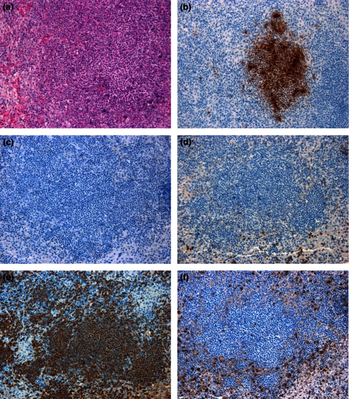
Figure 7.

Representative spleen sections from marmosets 48 h following inhalational challenge with Burkholderia pseudomallei HBPUB10303a. (a) Histological lesion (20×), (b) B. pseudomallei antigen stained brown (20×), (c) nitric oxide synthase expressing cell marker stained brown (20×), (d) B‐cell marker stained brown (20×), (e) macrophage/neutrophil stained brown, (f) T‐cell marker stained brown (20×).
