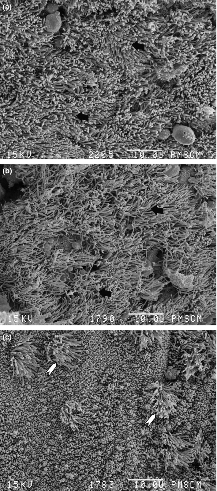
Figure 2.

Scanning electron microscopy image. No biofilm structure can be seen. (a) Patient no. 9 with normal epithelium and cilia, black arrow – cilia; (b) patient no. 3 with normal epithelium and cilia, black arrow – cilia; (c) patient no. 2, non‐ciliated cylindrical epithelium with few spots of cilia, white arrow head – cilia spots. Magnification 2000×.
