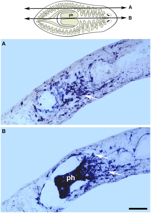
Figure 3.

Gtmhc expression in the regenerating pharynx. The cartoon represents the level and orientiation of the sagittal sections in (A,B). In situ hybridizations for a myosin heavy chain gene on sagittal sections of regenerating tails from the species Girardia tigrina, 6 days after amputation. (A) Lateral section showing an accumulation of myocytes (arrow) in the mesenchyme surrounding the pharynx. (B) Central section containing the regenerated pharynx (ph) with myocytes evident in the mesenchyme at its base (arrows). Scale bar: 50 μm. Image adapted from Cebrià (2000). Anterior to the right. Dorsal to the top.
