Figure 4.
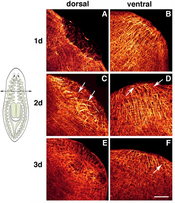
Planarian body-wall muscle regeneration. Whole-mount immunostaining with TMUS-13 antibody during head regeneration. Planarians were amputated at a pre-pharyngeal level as indicated in the cartoon. Head regeneration from the trunk piece was monitored. (A,B) Show dorsal and ventral views, respectively, of 1-day regenerants. A dorsal “hole” lacking fibers is evident in (A). (C,D) Show dorsal and ventral views, respectively, of 2-day regenerants. Pre-existing dorsal longitudinal fibers enter the blastema (arrows in C). The ventral region of the blastema contains mainly longitudinal fibers (arrows in D). (E,F) Show dorsal and ventral views, respectively, of 3-day regenerants. Dorsally, the muscle fibers converge at the center of the blastema, restoring the pattern observed in intact planarians. Ventrally, new circular muscle fibers are evident (arrow). Scale bar: 50 μm. Image adapted from Cebrià and Romero (2001).

