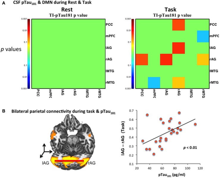
Figure 3.
CSF pTau181 level and functional connectivity during DMN. (A) CSF pTau181 during rest and task at various significant p-values; the significant p-values (y-axis) of the correlation between pTau181 and connectivity of brain regions were found only during task, but not during resting state; (B) pTau181 and bilateral inferior parietal connectivity during task. Individuals with stronger lAG–rAG have higher level of pTau181, which indicates higher risk of tau-related neural degeneration.

