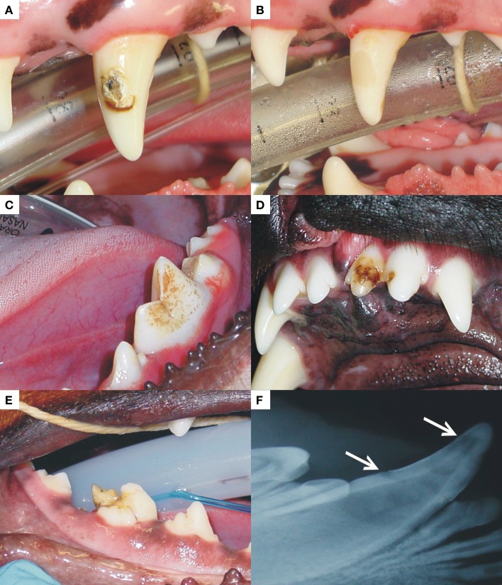
Figure 2.
A left maxillary canine (A) and left mandibular first molar (C) present with FEH, affecting the labial/buccal surfaces, respectively. The photograph in (D) is an example of FEH affecting two adjoining maxillary incisor teeth and (E) a right mandibular first molar with enamel hypoplasia that is so extensive that the distal cusp is completely distorted. The radiograph in (F) represents the enamel changes seen in a case of FEH. Radiographic changes may be so subtle that they can go unnoticed. The photograph marked as (B) was taken after restoration with a dental compomer of the tooth shown in (A).

