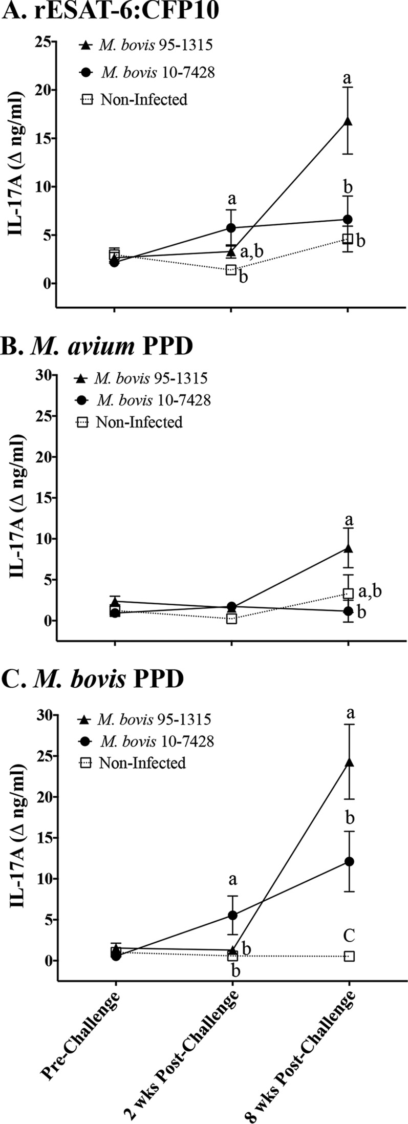
FIG 2.
IL-17A responses (protein) to M. bovis infection of cattle. Treatment groups included noninfected (n = 7), strain 95-1315-infected (white-tailed deer M. bovis isolate; n = 8), and strain 10-7428-infected (Holstein M. bovis isolate; n = 8) calves, with the experimental design described in Table 1. Whole blood was collected into heparinized tubes and stimulated with 1 μg/ml rESAT-6:CFP10 (A), 20 μg/ml M. avium PPD (Lelystad; Prionics Ag) (B), 20 μg/ml M. bovis PPD (Lelystad; Prionics Ag) (C), or medium alone (no stimulation) for 16 h at 39°C. Plasma was harvested for IL-17A analysis by ELISA (bovine IL-17A ELISA VetSet; Kingfisher Biotech). Data (mean ± SEM) are presented as the change in nanograms per milliliter (i.e., antigen stimulation minus medium alone) for each treatment group at the indicated time points relative to challenge. a to c, different letters indicate that responses differ for the given time point (P < 0.05, ANOVA followed by Tukey's multiple-comparison test).

