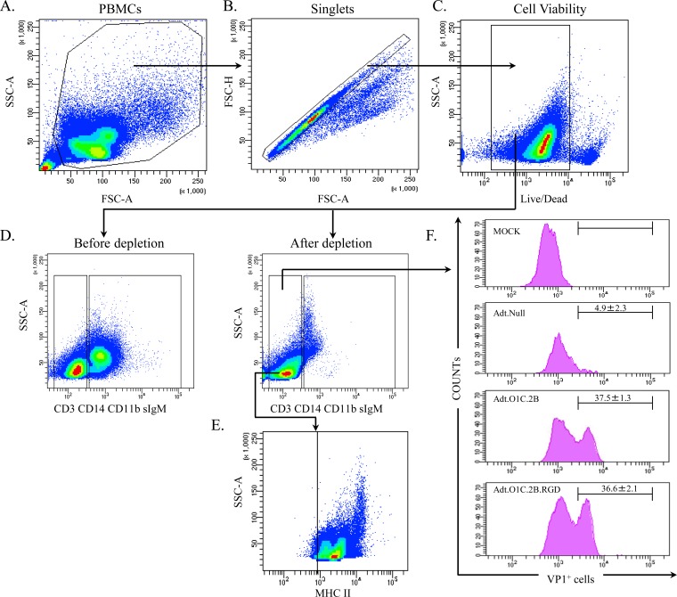
FIG 5.
Infection of the enriched APC population with Adt.O1C.2B or Adt.O1C.2B.RGD. (A) PBMCs from cows were subjected to an APC enrichment process. (B and C) Doublets (B) and dead cells (C) were excluded from the total PBMC population. (D) Using lineage-specific antibodies (anti-CD3, anti-CD14, anti-IgM, and anti-CD11b), T cells, monocytes, B cells, and NK cells were excluded. (E) The APC-enriched population was characterized using anti-MHC II antibody. (F) Lineage-negative cells were then infected with Adt.O1C.2B, Adt.O1C.2B.RGD, or Adt.Null at an MOI of 1 or were mock infected. Histograms show CD3-, CD11b-, CD14-, and IgM-negative cell populations that were stained for the VP1 FMDV structural protein. Data are representative of three independent experiments. Numbers on histograms, average percentages of cells expressing VP1 staining. SSC-A, side-scattered area; FSC-A, forward-scattered area; FSC-H, forward-scattered height.

