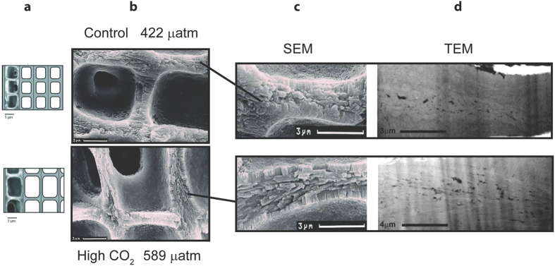
Figure 4.
Structural comparison of L. glaciale cell wall grown under natural conditions (top) and cultured under high CO2 conditions (bottom) using secondary electron microscopy (SEM, middle) (b,c) and transmitted electron microscopy (d) (TEM, right). The scale model (a) of L. glaciale on the far left is a modification of Ragazzola et al. (2012). Note the TEM images are not at the same scale. The higher CO2 growth results in thinner walls and larger cells due to the COs fertilisation of the photosynthesis (a,b). The control sample exhibits a narrow central channel structure, with low porosity and small crystallites with little alignment. In contrast the acidified sample shows strongly oriented, elongate crystals filling the central interstitial zone approximately parallel to the wall surface.

