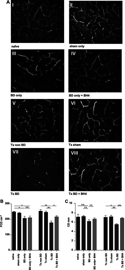
Figure 2.

Pancreatic microcirculation is disturbed following BD and IRI, but significantly improved following BH4 treatment (A) Fluorescein‐labeled dextran was used to visualize the capillary mesh of nontransplanted organs from: naïve animals (I), sham‐treated animals (II), BD donors (III), and BD donors treated with BH4 (IV). After syngeneic transplantation of pancreata derived from non‐BD donors into recipients an almost physiological capillary mesh was detected (V). Sham‐treated grafts displayed a regular capillary mesh after transplantation (VI). Severe microcirculatory alterations were detected after transplantation of untreated pancreata from BD donors (VII). In contrast, an amelioration of capillary graft microperfusion in the BH4 treatment group was observed (VIII). Representative pictures are shown for n = 5 animals/group. Observation area: 0.00159 cm2 (454 µm × 349 µm). (B) FCD defined as the length of all blood cell‐perfused capillaries per observation area. (C) CD defined as the mean of the three largest capillaries per observation area. Data are presented as mean of n = 5 animals/group ± SEM. Statistically significant differences were tested with one‐way ANOVA and the Tukey posttest; *p < 0.05, **p < 0.01, ***p < 0.001. ANOVA, analysis of variance; BD, brain dead; BH4, tetrahydrobiopterin; CD, capillary diameter; FCD, functional capillary density; IRI, ischemia reperfusion injury; SEM, standard error of the mean; Tx, transplantation.
