
Figure 5.
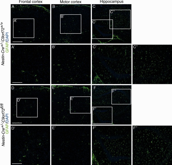
Immunostaining for glial fibrillary acidic protein (GFAP) in the brain of 18‐month‐old mice. Immunofluorescent staining is shown for GFAP in frontal cortex, motor cortex, and hippocampus of Nestin‐Cre+/−;C9orf72+/+ mice (A–C″) and Nestin‐Cre+/−;C9orf72fl/fl mice (D–F″). Boxes in A–C and D–F are shown at higher magnification in A′–C″ and D′–F″, respectively. No changes in the distribution, number, or appearance of GFAP‐positive astrocytes were detected in Nestin‐Cre+/−;C9orf72fl/fl mice. Scale bars: A, D, 200μm; A′, D′, 100μm. DAPI = 4′,6‐diamidino‐2‐phenylindole.