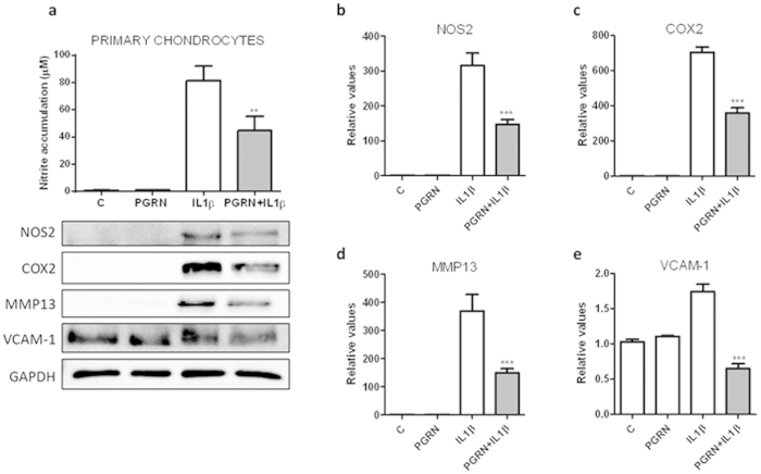
Figure 4. PGRN suppressed IL1-β induction of chondrocyte catabolism.
(a) Cells were treated with 10 ng/ml of IL1β, in presence or absence of 200 ng/ml of PGRN for 48 h. Culture medium was subsequently analyzed for nitrite levels. NO concentration (μM) was determined using the Griess reaction. Values are the mean ± SEM of at least 3 independent experiments (**P < 0.01 vs. IL1β) (a, upper panel). Cell lysates underwent Western blotting analysis under the same condition (a, lower panel). After transferring the blots onto PVDF membranes, we cropped the targeted blots according to referenced indicating markers, and then targeted proteins were immunoblotted with NOS2, COX-2, MMP13 and VCAM-1 antibodies. GAPDH was used as a loading control. Blots are representative of at least 3 independent experiments. (b–e) Western blot densitometric analysis (n = 3; ***P < 0.001 PGRN + IL1β vs. IL1β).

