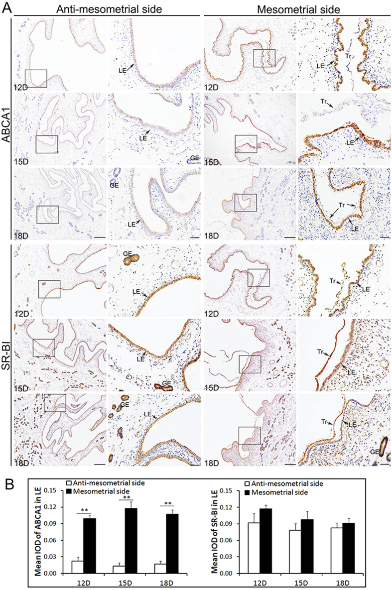
Figure 2. Immunohistochemical analysis of ABCA1 and SR-BI at the maternal-fetal interface (including the anti-mesometrial side and mesometrial side of the uterus) during the peri-implantation period in Yorkshire pigs.
The sections stained with isotype matched normal rabbit IgG served as negative control (the control images of ABCA1 and SR-BI were in the Figs 5 or 7). (A) Images stained with ABCA1 and SR-BI antibodies on Days 12, 15 and 18 of pregnency in Yorkshire pigs. The results show that the ABCA1 signals were observed mainly to LE at the mesometrial side and were barely detected in LE at the anti-mesometrial side. The positive signals were also barely detected in Tr and GE. The SR-BI signals were observed in LE, GE and Tr. The stain intensity of SR-BI in LE between at the mesometrial side and the anti-mesometrial side was similar. (B) Quantitative analysis of ABCA1 and SR-BI by measuring the average integrated optical density (IOD) in LE in Yorkshire pigs. Asterisks indicate significant differences (mean ± SD) between breeds (**P < 0.01), P value was determined by Student’s t test. Legend: Tr, trophoblast; LE, endometrial luminal epithelium; GE, glandular epithelium; D, day of pregnancy; Scale bars = 100 μm or 20 μm (amplification).

