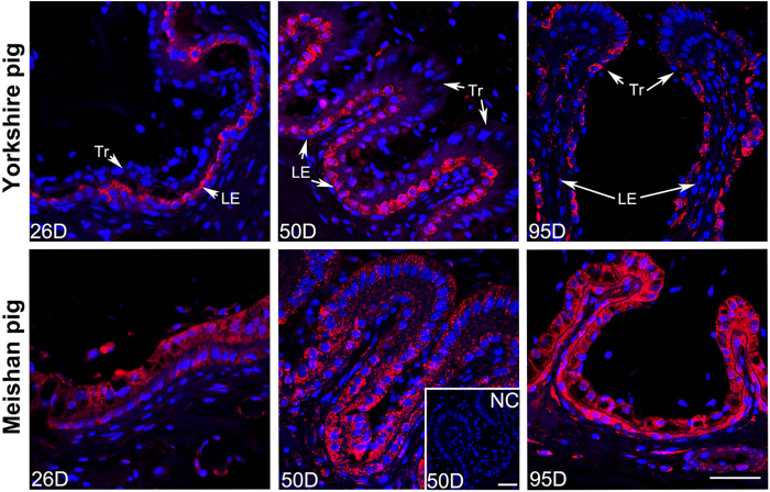
Figure 6. Confocal laser scanning microscopic localization of ABCA1 using rabbit anti-human ABCA1 polyclonal antibody at the maternal-fetal interface in Yorkshire and Meishan pigs.
Positive staining of ABCA1 was shown in red and the staining was diffused in the positive cells (except the nuclei) without specific subcellular localization. The blue staining represents nuclei (DAPI stained). In Yorkshire pigs, the ABCA1 staining were mostly observed in LE on Days 26 and 50 of pregnancy, while on Day 95 of pregnancy, the positive signals were mostly observed in Tr and just a few positive signals could be observed in LE. In Meishan pigs, the ABCA1 staining was observed both in Tr and LE on Days 26, 50 and 95 of pregnancy. Legend: Tr, trophoblast; LE, endometrial luminal epithelium; GE, glandular epithelium; D, day of pregnancy; NC, negative control; Scale bar = 40 μm.

