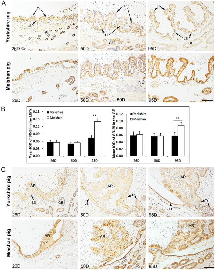
Figure 7. Immunohistochemical analysis of SR-BI at the maternal-fetal interface and the placental areolae in Yorkshire and Meishan pigs.
Uterine/placental sections were obtained on Days 26, 50 and 95 of pregnancy. Tissue sections were stained with rabbit anti-human SR-BI polyclonal antibody. The sections stained with isotype matched normal rabbit IgG served as negative control. (A) Images were taken from the maternal-fetal interface in Yorkshire and Meishan pigs. SR-BI positive cells were observed in Tr, LE and GE in Yorkshire and Meishan pigs on Days 26, 50 and 95 of pregnancy. Legend: Tr, trophoblast; LE, endometrial luminal epithelium; GE, glandular epithelium; D, day of pregnancy; NC, negative control; Scale bar = 100 μm. (B) Quantitative analysis of SR-BI by measuring the average integrated optical density (IOD) in the epithelial bilayer (composed by Tr and LE) and GE during pregnancy. Asterisks indicate significant differences (mean ± SD) between breeds within a day (**P < 0.01. Analyzed by PROC MIXED of SAS). The data were shown as the mean ± SD. (C) Images were taken from the placental areolae in Yorkshire and Meishan pigs. The SR-BI positive signals were filled with all the areolar regions, including Tr, LE and GE on Days 26, 50 and 95 of pregnancy. AR, areola ; Scale bar = 100 μm.

