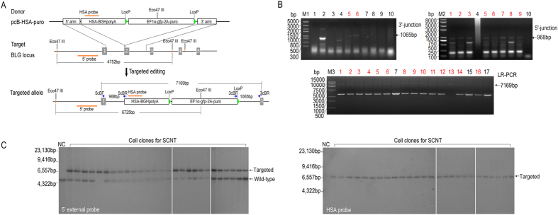
Figure 4. HSA gene targeting at BLG locus by TALE nickase-mediated HDR in BFFs.
(A) Schematic of the targeting strategy for BLG locus. Southern blot probes and primers for PCR detection are shown as red lines and blue arrows, respectively. Gray boxes indicate the exons of BLG. HSA-BGHpolyA, HSA encoding sequence followed by BGHpolyA; and EF1α-gfp-2A-puro, elongation factor 1α promoter followed by GFP gene, a 2A self-cleaving peptide sequence and puromycin resistance gene. (B) PCR detection of drug-resistant cell clones. (Top) Left image shows the results of the 3′ junction PCR performed on drug-resistant cell clones using the primers 3cBF/R, and the right image shows the results of the 5′ junction PCR performed on 3′ junction- positive cell clones using primers 5cBF/R. (Bottom) Results of the LR-PCR performed on the 5′ junction positive cell clones using the primers 5cBF/3cBR. Lane 13 shows the bi-allelic targeted cell clone; the expected PCR products are shown by arrows. The red numbers indicate the PCR-positive clones. (C) Results of Southern blot conducted on cell clones with normal chromosome. 5′-probe (left) detected a 6.73 kb targeted fragment and 4.75 kb wild type fragment; HSA probe (right) detects a 6.73 kb targeted fragment.

