Fig. 2.

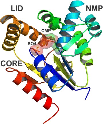
Ribbon diagram of the structural model of M. tuberculosis cytidilate monokinase (Rv1712). The CORE domain, NMP-binding domain and the LID domain are labelled as well as ligands CMP and SO4 shown as sticks. Figure generated using PyMOL [45]

Ribbon diagram of the structural model of M. tuberculosis cytidilate monokinase (Rv1712). The CORE domain, NMP-binding domain and the LID domain are labelled as well as ligands CMP and SO4 shown as sticks. Figure generated using PyMOL [45]