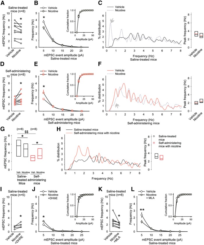
Figure 7.
Nicotine in vitro blocks CPD through α7*- and α4β2*-type nicotinic receptors. A, Responses of individual MSNs show that nicotine inhibited corticostriatal activity in saline-treated mice by (B) reducing high-frequency, low-amplitude mEPSCs, but had no effect on their amplitude distribution (inset). For B, E, J, *p<0.05, Bonferroni t test; for all other panels, *p<0.05, paired t test and $$p<0.01, Student’s t test. C, The frequency distribution (left) and the average peak frequency (right) of mEPSCs. Nicotine increased low-frequency mEPSCs in saline-treated mice. D, Nicotine increased the frequency of mEPSCs in MSNs from amphetamine self-administering mice by (E) augmenting high-frequency, low-amplitude mEPSCs, while having no effect on their amplitude distribution (inset). F, The frequency distribution (left) and average peak frequency (right) of mEPSCs. Nicotine reduced low-frequency mEPSCs in amphetamine self-administering mice. G, Mean±SE frequencies of mEPSCs from saline-treated and amphetamine self-administering mice in response to nicotine. H, The frequency distribution (left) and average peak frequencies (right) compares mEPSCs in MSNs from saline-treated mice with those from amphetamine self-administering mice exposed to nicotine in vitro. I, The α4β2*-type receptor antagonist DHβE blocked the synaptic depression generated by nicotine and (J) increased high-frequency, low-amplitude synaptic events. K, The α7*-type nicotinic receptor antagonist MLA reduced (L) high-frequency, low-amplitude mEPSCs. J, L, Insets show similar mEPSC amplitude distributions.

