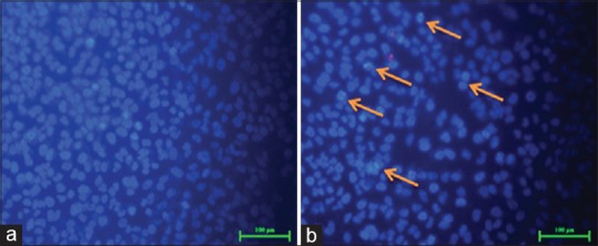
Figure 6.

4’, 6-diamidino-2-phenylindole staining shows the presence of blue fluorescent cells in Cissus quadrangularis extract treated ones shown by arrows in the Figure b whereas low fluorescence in control section (a) control. However the number of cells also decreased after the Cissus quadrangularis extract treatment (b)
