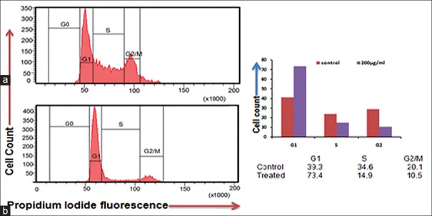
Figure 8.
Cissus quadrangularis extract induced apoptosis in KB cells assayed by flow cytometry by propidium iodide staining. Cells were treated with 200 μg/ml of Cissus quadrangularis for 24 h, stained by propidium iodide and followed by flow cytometry to determine the hypodiploid DNA (fragmented DNA) proportions. The percentage of hypodiploid cells (sub G1 peak) was calculated on the basis of the respective histograms. (a) Untreated cells, (b) 200 μg/ml Cissus quadrangularis treated KB cells. Respective histogram shows G1 phase arrest in treated ones as compares to control

