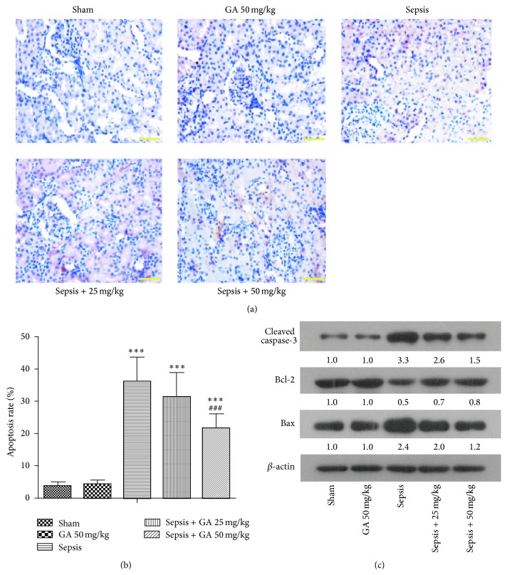
Figure 5.
GA inhibited the apoptosis induced by AKI in kidney tissue. (a) The apoptosis of cells in kidney tissue was detected by TUNEL staining (magnification 400x). (b) The apoptosis rate of TUNEL staining from different groups was calculated. (c) Apoptosis-related proteins were detected by western blot. β-actin was used as a loading control. The results shown are representative of at least three independent experiments. Each value represents the mean ± SD (n = 6). ∗∗∗ P < 0.001, versus the sham operation group. ### P < 0.001 versus the sepsis group.

