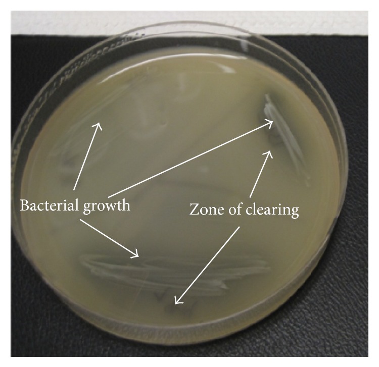
Figure 4.

Phenotypic detection of hyaluronidase production. Plates were prepared with brain heart infusion agar supplemented with hyaluronic acid and albumin. P. acnes isolates were allowed to grow until there was visible growth; thereafter, the plate was acidified with 2 N glacial acetic acid, causing undigested hyaluronic acid to bind to albumin, forming a white precipitate. Hyaluronidase production was considered to be present if a zone of clearing was observed.
